|
|
Broncholithiasis
Broncholiths
General Considerations
- Uncommon
- Calcification within a bronchus, most commonly from peribronchial nodes that calcify following inflammation
- If adjacent to a bronchus, calcified node may erode into the lumen forming a broncholith
- Usually associated with prior tuberculosis or histoplasmosis
- Some authors have expanded definition to include peribronchial calcification as well
- Rarely may be caused by aspiration of previously calcified object, like a bone fragment
- Bronchoscopy may fail to detect the calcification because it is obscured by the inflammatory process surrounding it
Clinical Findings
- Non-productive cough
- Hemoptysis
- Pain
- Chills and fever from a secondary infection
- Lithoptyis-expectoration of calcified material
Imaging Findings
- Chest radiography usually does not show the calcification or its exact location
- Helical CT is the imaging study of choice
- The right middle lobe bronchus and the anterior segmental bronchi of the upper lobes are favored sites
- Calcified endobronchial or peribronchial lymph node
- No associated soft tissue mass
- Atelectasis is the most common parenchymal finding
- Mucoid impaction
- Post-obstructive infection may result
- Bronchiectasis
- Expiratory air-trapping is uncommon
Differential Diagnosis
- Carcinoid tumor
- Calcification in a fungus ball
- Hamartomas
Treatment
- Symptomatic broncholiths may be surgically removed
- Bronchoscopic attempt at removal of a broncholith still embedded in wall may result in hemorrhage
Complications
- Rarely, bronchoesophageal or broncho-aortic fistulae
 
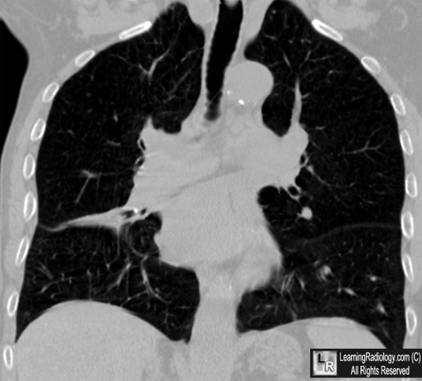
Broncholithiasis. Left: Coronal reconstruction of CT of the chest demonstrates a
calcified
broncholith (white arrow) within the right middle lobe bronchus.
It is producing partial atelectasis
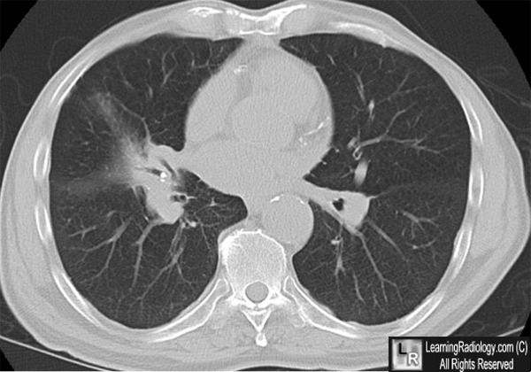
of the right middle lobe (yellow arrow). Right: Axial CT image demonstrates broncholiths
within bronchus (white arrow) with associated parenchymal disease.
For these same photos without the arrows, click here and here
For more information, click on the link if you see this icon 
Broncholithiasis: CT Features in 15 Patients. DJ Conces, Jr.; RD Tarver and VA Vix. AJR 157:249-253, August 1991
Broncholithiasis: Review of the Causes with Radiologic-Pathologic Correlation. JB Seo, Koun-Sik Song, JS Lee, JM Goo, HY Kim, Jae-Woo Song, IS Lee, and Tae-Hwan Lim. RadioGraphics 2002; 22:S199–S213
|
|
|
